5.1 CNS Regulation, Mood, and Cognition Concepts V2
Concepts Related to the CNS, Mood, and Cognition
This resource provides a basic introduction to the concepts related to the central nervous system and cognition in connection with pharmacology. For a more detailed exploration of the central nervous system, refer to an anatomy and physiology resource.
The concept of cognition is defined as “the process of thought that embodies perception, attention, visuospatial cognition, language, learning, memory, and executive function with the higher-order thinking skills of comprehension, insight, problem-solving, reasoning, decision making, creativity, and metacognition” (Giddens, 2017).
Jessica/ Jan 27: @Copyeditor the numbered hyperlinks (such as [2], [3], and [4]) are currently directing me to Version 1 of the book on BCcampus.
This chapter also addresses mood[2], affect[3], and anxiety[4] (Giddens, 2017).
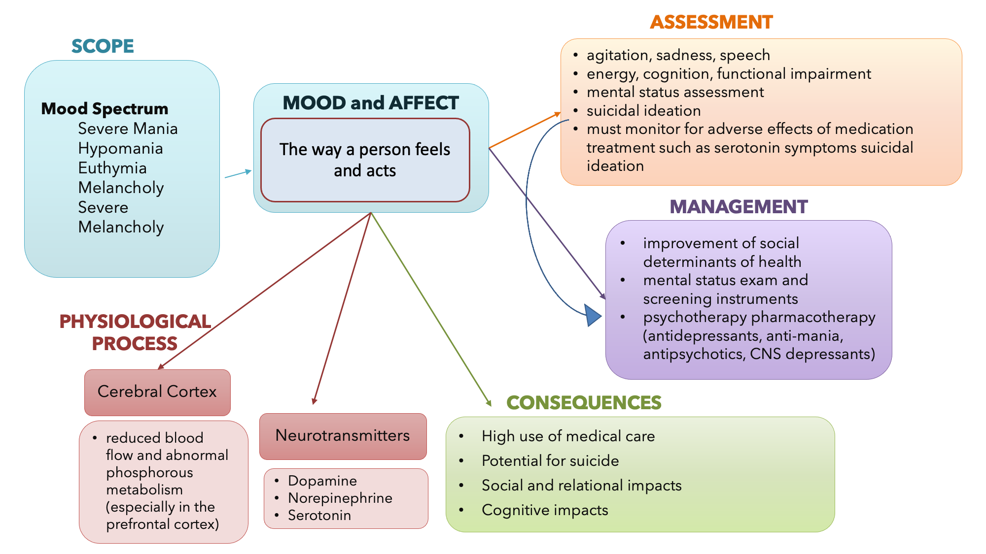
The concept map in Figure 8.2a summarizes information related to the concept of mood, affect, and the CNS. You are encouraged to revisit this map after you have completed the chapter. You may also wish to develop your own concept map related to other CNS concepts in this chapter.
Overview of the Central Nervous System and Processes
Before we can begin to understand how different medications influence the brain, we need to review the central nervous system. The nervous system can be divided into two major regions: the central and peripheral nervous systems. The central nervous system (CNS) is the brain and spinal cord, and the peripheral nervous system (PNS) is everything else. The brain is contained within the cranial cavity of the skull, and the spinal cord is contained within the vertebral cavity of the vertebral column.
It is a bit of an oversimplification to say that the CNS is what is inside these two cavities and the peripheral nervous system is outside of them, but that is one way to start to think about it. In actuality, there are some elements of the peripheral nervous system that are within the cranial or vertebral cavities. The peripheral nervous system is so named because it is on the periphery—meaning beyond the brain and spinal cord. Depending on different aspects of the nervous system, the dividing line between central and peripheral is not necessarily universal.
Brain
The brain is a highly complex organ with over 86 billion neurons that communicate between each other and to other parts of the body. The brain acts as a command center with numerous functions including processing sensory information, regulating body functions, controlling movement and overseeing cognition (The Society of Neuroscience, nd).
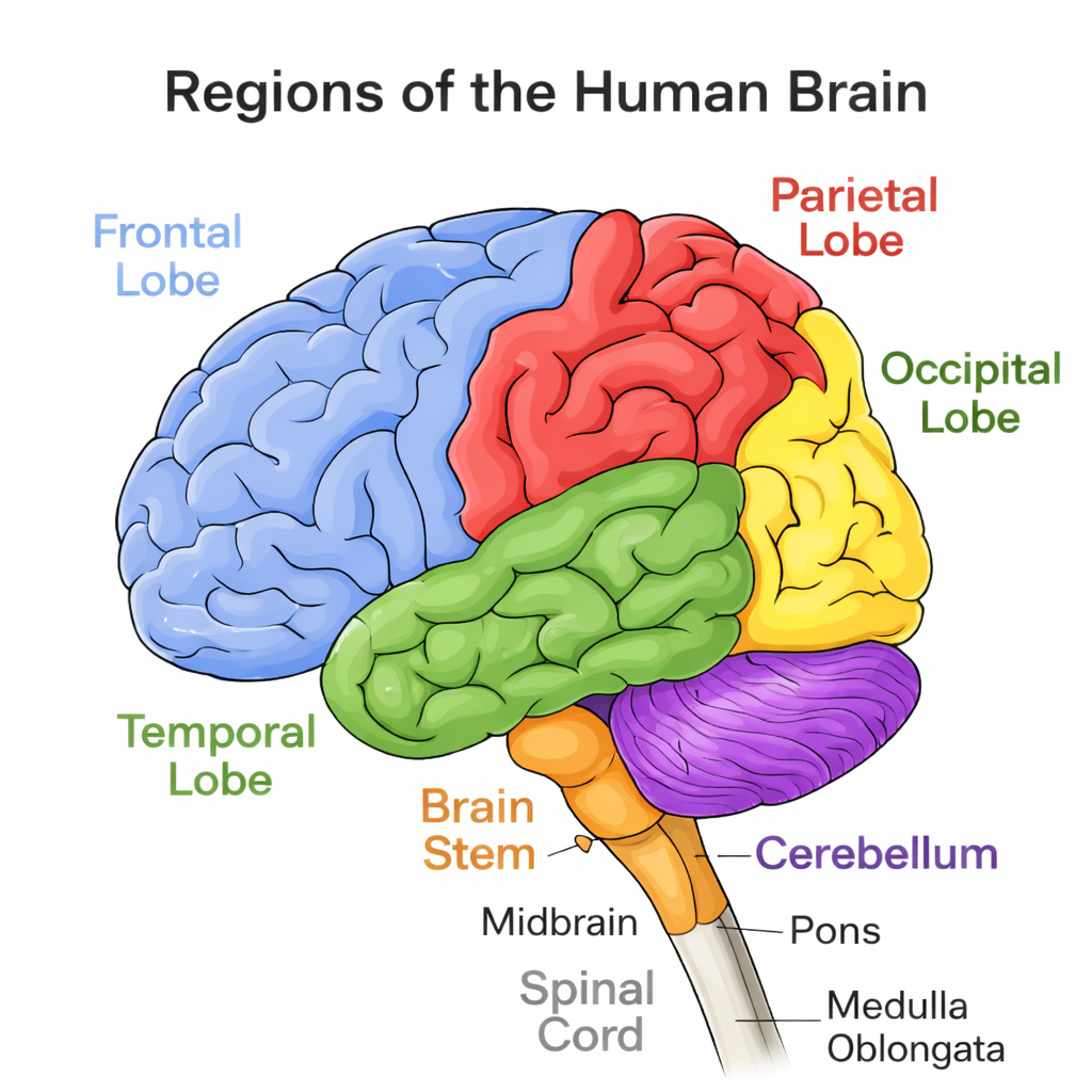
The brain is divided into different regions with each region designed to do special tasks or abilities. Basic brain structures and their functions are listed below (The Society of Neuroscience, nd; Adams et al, 2018).
| Anatomy of the Brain | Function |
| Cerebrum: two large hemispheres containing cerebral cortex and corpus callosum. | Carry information from one side of the brain to the other parts.
“thinking” part of the brain, responsible for perception, speech, motor movements, memory and smell. |
| Cerebral Cortex: within the cerebrum, divided into lobes | Frontal lobe: coordinate voluntary movements and speech, memory and emotion, complex thought (ie. planning and problem-solving), and many aspects of personality (moral and ethical behaviour).
Parietal: integrate sensory signals from the skin, process taste, and process some types of visual information. Occipital: process visual information Temporal: hearing, equilibrium, emotion and memory (amygdala), visual processing. |
| Thalamus | integrates sensory information and relays it to other parts of the brain, |
| Hypothalamus | which sends hormonal signals to the rest of the body through the pituitary gland. |
| Cerebellum | coordinates voluntary movements and helps the brain learn new motor skills. It also has roles in spatial and temporal perception |
| Brainstem: includes midbrain, pons and medulla oblongata. | Pons: influences breathing and posture
Midbrain: motor control (especially eye movement), auditory and visual processing, and regulating alertness, sleep, and pain; it is a key relay center, connecting sensory and motor pathways between the forebrain and the rest of the brainstem. medulla: carries nerve pathways connecting the brain to the spinal cord and contains neural networks that help control basic functions like swallowing, heart rate, and breathing. |
Watch the supplementary video explaining the brain and its function.
Video 5.1c. Neuroscientifically Challenged (April 4, 2025). Major Brain Structures and their Functions. Major Brain Structures and Their Functions
The Peripheral Nervous System
The peripheral nervous system is further divided into the autonomic nervous system and the somatic nervous system, which are further discussed in the Autonomic Nervous System chapter in this book. (See Figures 5.1d and 5.1e for illustrations of the central and peripheral nervous systems).


Review more detailed information about the nervous system function using this OpenStax link: Basic structure and function of the nervous system
Communication in the Nervous System
Neurons are the primary cells involved in processing information. There are thousands of individual cell types based on morphology, location, connectivity and chemistry (Hyman, 2005). Neurotransmitters (NTs) are chemical substances that carry information between the neurons and they play a central role in brain function and are involved in receiving, processing and transmitting information. Your brain communicates with electrical impulses that signal a release of a neurotransmitter, which then binds to the targeted cell.
Understanding this communication will help you put the pieces together when you are trying to understand the mechanism of action of a medication that works by influencing neurotransmitters.
See Figure 5.1e for an illustration of the major elements in neuron communication.
/10%3A_Overview_of_the_Nervous_System/10.1%3A_Introduction_to_the_Nervous_System/10.1A%3A_Organization_of_the_Nervous_System")
There are two types of connections between electrically active cells: chemical synapses and electrical synapses. In a chemical synapse, a chemical signal—namely, a neurotransmitter—is released from one cell and affects another cell. In comparison, in an electrical synapse, there is a direct connection between the two cells so that ions can pass directly from one cell to the next. In this unit, we will be focusing on the communication of a neurotransmitter in a chemicalsynapse. Once in the synaptic cleft, the neurotransmitter diffuses the short distance to the postsynaptic membrane and can interact with neurotransmitter receptors. Receptors are specific for the neurotransmitter, and the two fit together like a key and lock. One neurotransmitter binds to its receptor and will not bind to receptors for other neurotransmitters, making the binding a specific chemical event (OpenStax, 2025). (See Figure 5.2e for an illustration of a synapse).
When the neurotransmitter binds to the receptor, the cell membrane of the target neuron changes its electrical state, and a new graded potential begins. If that graded potential is strong enough to reach threshold, the second neuron generates an action potential. The target of this neuron is another neuron in the thalamus of the brain, the part of the CNS that acts as a relay for sensory information. The thalamus then sends the sensory information to the cerebral cortex, the outermost layer of gray matter in the brain, where conscious perception of that stimulus begins (OpenStax, 2025).
Neurotransmitters simplified
A signal is sent: an electrical impulse (action potential) travels down a neuron.
Neurotransmitters released: this triggers chemical messengers to be released into the synapse.
Message received: these messengers bind to receptors on the next neuron.
The signal is passed on or stopped: the receiving neuron is either activated (excited), calmed (inhibited), or influenced in other ways.
Enzyme clean up: any leftover neurotransmitters are either broken down by enzymes or taken back up by the original neuron (reuptake).
Guy-Evans, 2025.

Chemical Synapse.jpg by Young et al., 2018 under OpenStax Anatomy and Physiology the Creative Commons Attribution 4.0 International license.
A supplementary video explaining neuron communication via action potentials is provided below.
Jessica/Jan 27: @Copyeditor please be aware that hyperlink below takes us to version 1 of the book where the footnotes are.
Neuron communication via Action Potentials[13]
Types of Neurotransmitters
There a number of different types of neurotransmitters. In the CNS, there are at least 21 neurotransmitters and likely more that have yet to be fully discovered. Some NTs are exclusive to the CNS, and others are in both the peripheral nervous system, autonomic nervous system or both. The response of the NT when released from the presynaptic cleft, crosses the synapse, and then either activates or inhibits the receptor on the postsynaptic neuron.
There are six groups of neurotransmitters. Biogenic Amines, amino acids, purines (ie. Adenosine), opioid peptides (ie. Endorphins, encephalins), and nonopioid peptides (ie. Oxytocin, substance P, vasopressin). Acetylcholine and histamine are not grouped within these categories. Biogenic amines and amino acids are explained in more detail below (Rosenjack Burchum & Rosenthal, 2019).
Amino Acids
One group of neurotransmitters is amino acids. GABA (gamma-aminobutyric acid) is an example of an amino acid neurotransmitter. They each have their own receptors and do not interact with each other. Amino acid neurotransmitters are eliminated from the synapse by re-uptake. A pump in the cell membrane of the presynaptic element, or sometimes a neighbouring glial cell, will clear the amino acid from the synaptic cleft so that it can be recycled, repackaged in vesicles, and released again.
Biogenic Amine (also known as monoamine)
Another class of neurotransmitters is the biogenic amine, a group of neurotransmitters that are enzymatically made from amino acids. For example, serotonin is made from tryptophan. It is the basis of the serotonergic system, which has its own specific receptors. Serotonin is transported back into the presynaptic cell for repackaging.
Other biogenic amines are made from tyrosine and include dopamine, norepinephrine, and epinephrine. Dopamine is part of its own system, the dopaminergic system, which has dopamine receptors. Norepinephrine and epinephrine belong to the adrenergic neurotransmitter system. The two molecules are very similar and bind to the same receptors, which are referred to as alpha- and beta-receptors. The biogenic amines have mixed effects. For example, dopamine receptors that are classified as D1 receptors are excitatory, whereas D2-type receptors are inhibitory.
Functions of Neurotransmitters
There are three main functions of neurotransmitters:
- Excitatory: Increases the chance of the neuron firing.
- Inhibitory: Calming effect, reduces the chance of firing.
- Modulatory: Adjust the activity of other neurotransmitters. Ie. Dopamine.
Excitatory Neurotransmitters
Excitatory Neurotransmitters |
Excitatory Neurotransmitters |
|
Acetylcholine (in CNS) Role in learning, memory Regulates mood, mania, sexual aggression Activates muscle action Decreased: Alzheimer’s Disease, Parkinson’s Disease Increased: schizophrenia |
Norepinephrine (noradrenaline) Role in mood, attention, arousal Fight or flight response to stress (stimulates SNS) Contracts blood vessels, increases blood flow Decreased: depression, loss of interest Increased: mania, anxiety, schizophrenia, addiction |
|
Adrenaline Similar to norepinephrine, but more hormone-like. Heightens alertness and prepares the body for action. Increased: high blood pressure, stress
|
Glutamate Main excitatory neurotransmitter in the brain Crucial for learning and memory Works with GABA to control many brain function including level of excitation & regulates action potential of cells. Decreased (NMDA): psychosis Increased (NMDA): neurotoxic, Alzheimer’s, stroke |
Inhibitory Neurotransmitters |
Inhibitory Neurotransmitters |
|
Gamma-aminobutyric acid (GABA) Main calming neurotransmitter Helps regulate anxiety, motor control, and sleep
Decreased: anxiety, seizures, mood disorders Increased: sedation, reduction of anxiety |
Dopamine Involved in pleasure, motivation, movement, and learning Stimulates hypothalamus to release hormones Acts as both excitatory and inhibitory, depending on receptor. Modulates affective states and emotions Decreased: Parkinson’s, depression Increased: schizophrenia, mania, ADHD |
|
Glycine (in CNS) Slows nerve signals, controls motor coordination, in brainstem and spinal cord Controls vision and pain perception
|
Serotonin Regulates mood, sleep, appetite, and digestion Decreased: depression, anxiety, insomnia Increased: anxiety, insomnia |
Guy-Evans, 2025; Halter et al, 2019
Neurotransmitters play such a central role in brain function, that an imbalance of NTs can lead to many psychiatric, neurologic, and physiological conditions. Treatment with medications for many conditions target neurotransmitter receptors and other proteins involved in neurotransmitter synthesis and inactivation. The medications that are used to treat CNS disorders mimic or block the neurotransmitter based on the imbalance caused by the condition. Medications are used to either stimulate or depress the effect of the neurotransmitter. For example, CNS depressants alter the brain by decreasing the excitability of neurotransmitters, blocking their receptor site, or increasing the inhibitory neurotransmitter. On the other hand, CNS stimulants increase brain activity by increasing the excitability of neurotransmitters, decreasing the inhibitory neurotransmitters, or blocking their receptor sites (Velarde, 2018).
Norepinephrine is often associated with the fight-or-flight response. Abnormal levels of this neurotransmitter are also associated with depression, decreased alertness and interest, along with possible palpitations, anxiety, and panic attacks. Dopamine is strongly linked to motor and cognition. This neurotransmitter influences movement and can be associated with ADHD, paranoia, and schizophrenia. Serotonin is heavily involved in many bodily processes. Abnormal levels of serotonin can affect sleep, libido, mood, and temperature regulation. Alterations of this neurotransmitter have been linked to many mental health issues such as depression, bipolar disorder, anxiety, and body disorders. GABA (gamma-aminobutyric acid) can act as an inhibitory neurotransmitter. GABA assists with communication in the brain, and if this neurotransmitter is low, it has been linked to issues such as anxiety, seizures, mania, and impulse control. The neurotransmitter glutamate works as an excitatory neurotransmitter and works with GABA to control other functions of the brain (Velarde, 2018).
Image Descriptions
Figure 5.2a Mood and Affect Concept Map
Mood and affect – the way a person feels and acts.
Scope – mood spectrum:
- severe mania
- hypomania
- euthymia
- melancholy
- severe melancholy
Physiological process
- Cerebral cortex
- reduced blood flow and abnormal phosphorous metabolism (especially in the prefrontal cortex)
- Neurotransmitters
- dopamine
- norepinephrine
- serotonin
Assessment
- agitation, sadness, speech
- energy, cognition, functional impairment
- mental status assessment
- suicidal ideation
- must monitor for adverse effects of medication treatment such as serotonin symptoms and suicidal ideation
Management
- improvement of social determinants of health
- mental status exam and screening instruments
- psychotherapy pharmacotherapy (anti-depressants, anti-mania, antipsychotics, CNS depressants).
Consequences
- high use of medical care
- potential for suicide
- social and relational impacts
- cognitive impacts
Figure 8.2c Somatic, Autonomic, and Enteric Structures of the Nervous System
Brain (CNS)
- perception and processing of sensory stimuli (somatic/autonomic)
- execution of voluntary motor responses (somatic)
- regulation of homeostatic mechanisms (autonomic)
Spinal cord (CNS)
- initiation of reflexes from ventral horn (somatic) and lateral horn (autonomic) gray matter
- pathways for sensory and motor functions between periphery and brain (somatic/autonomic)
Nerve (PNS)
- fibers of sensory and motor neurons (somatic/autonomic)
Ganglia (PNS)
- reception of sensory stimuli by dorsal root and cranial ganglia (somatic/autonomic)
- relay of visceral motor responses by autonomic ganglia (autonomic)
Digestive tract (ENS)
- the enteric nervous system (ENS), located in the digestive tract, is responsible for autonomous functions and can operate independently of the brain and spinal cord.
Images:
- 5.1a Mood and Affect Concept Map [Image description]
- 5.1b Lobes of the brain identified (Sheila Odubote/ TRU Open Press)
- 5.1c “1201 Overview of Nervous System.jpg” by OpenStax is licensed under CC BY 4.0. Access for free at https://openstax.org/books/anatomy-and-physiology/pages/12-1-basic-structure-and-function-of-the-nervous-system ↵
- 5.1d “1205 Somatic Autonomic Enteric StructuresN.jpg” by OpenStax is licensed under CC BY 4.0. Access for free at https://openstax.org/books/anatomy-and-physiology/pages/12-1-basic-structure-and-function-of-the-nervous-system ↵
- 5.1e “Chemical synapse schema cropped.jpg” by Looie496 is licensed under public domain. Access for free at https://med.libretexts.org/Bookshelves/Anatomy_and_Physiology/Book%3A_Anatomy_and_Physiology_(Boundless)/10%3A_Overview_of_the_Nervous_System/10.1%3A_Introduction_to_the_Nervous_System/10.1A%3A_Organization_of_the_Nervous_System ↵
- “1225 Chemical Synapse.jpg” by Young, KA., Wise, JA., DeSaix, P., Kruse, DH., Poe, B., Johnson, E., Johnson, JE., Korol, O., Betts, JG., & Womble, M. is licensed under CC BY 4.0 Access for free at https://openstax.org/books/anatomy-and-physiology/pages/12-5-communication-between-neurons ↵
- Adams, M., Urban, C., El-Hussein, M., Osuji, J. & King, S. (2018). Pharmacology for Nurses. A pathophysiological approach (2nd Canadian ed.). Pearson Canada Inc: Ontario.
- Hyman, S. (2005). Neurotransmitters. Current Biology, 15(5), R154 – R158
- Forciea, B. (2015, May 12). Anatomy and Physiology: Nervous System: Action Potential Generation V2.0. [Video]. YouTube. All rights reserved. Video used with permission. https://youtu.be/-xFliVq3MKg. ↵
- Giddens, J. (2017). Concepts of Nursing Practice (2nd ed.). Missouri: Elsevier, pg. 319.
- Guy-Evans, O. (2025). Neurotransmitters: types, functions, examples. Simply Psychology.https://www.simplypsychology.org/neurotransmitter.html
- Halter, M., Pollard, C. & Jakubec, S. (2019). Varcarolis’s Canadian Psychiatric Mental Health Nursing. A clinical approach (2nd ed.). Elsevier: Canada
- OpenStax (2025). Anatomy and Physiology licensed under CC BY 4.0. https://openstax.org/books/anatomy-and-physiology/pages/1-introduction ↵
- Rosenjack Burchum, J. & Rosenthal, L. (2019). Lehne’s pharmacology for nursing care (10th ed.). Elsevier: Canada
- The Society of Neuroscience (2018). Brain Facts. The Brain Facts Book
- Velarde, G. (2019). Pharmacology Notes: Nursing Implications for Clinical Practice licensed under CC BY-NC-SA 4.0. ↵
the process of thought that embodies perception, attention, visuospatial cognition, language, learning, memory, and executive function with the higher order thinking skills of comprehension, insight, problem solving, reasoning, decision making, creativity, and metacognition
the way a person feels [1].
the observable response a person has to his or her own feelings, [2]
an alert to the human condition of impending doom, either real or imagined, and is accompanied by autonomic responses that serve as protective [3].
Anatomical division of the nervous system located within the cranial and vertebral cavities, namely the brain and spinal cord.
An anatomical division of the nervous system that is largely outside the cranial and vertebral cavities, namely all parts except the brain and spinal cord.
Cells that carry electrical impulses to the synapse of a target organ.
Connection between two neurons, or between a neuron and its target, where a neurotransmitter diffuses across a very short distance.
Connection between two neurons, or any two electrically active cells, where ions flow directly through channels spanning their adjacent cell membranes.
The membrane voltage at which an action potential is initiated.
A change in voltage of a cell membrane in response to a stimulus that results in transmission of an electrical signal; unique to neurons and muscle fibers.
The region of the central nervous system that acts as a relay for sensory pathways.
