9.2 Overview of the GI System
It is important to understand the GI anatomy and physiology to understand how the GI medications work to treat conditions. The GI system is complex and this unit will provide a general overview including digestive system structure, processes and regulation.
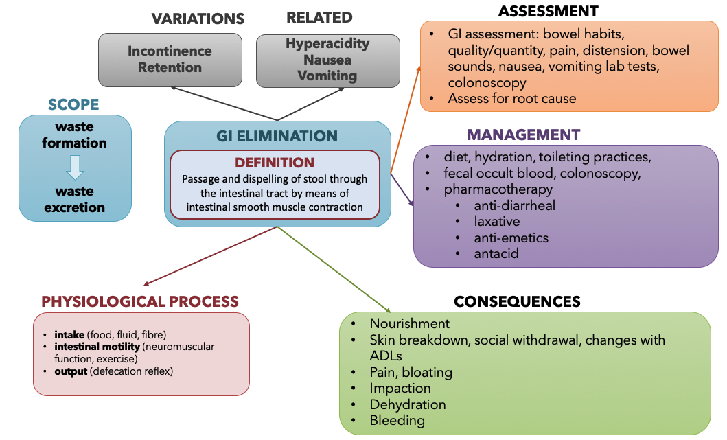
The example concept map below in figure 9.2a provides a summary of the key information necessary to understand gastrointestinal elimination informed by several resources (Giddens, 2017).
You are encouraged to revisit this map after you have completed the chapter.
Overview of Gastrointestinal System and Processes
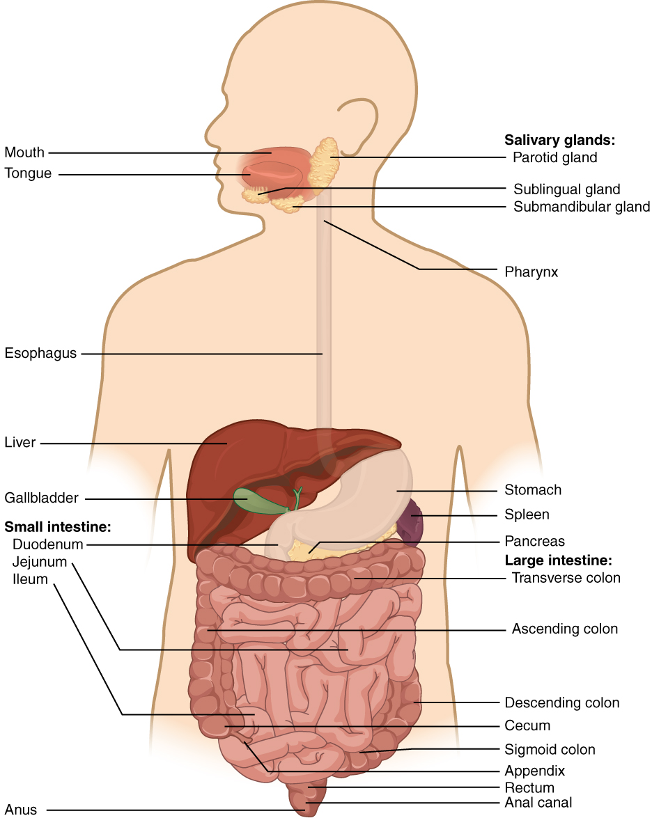
The digestive system has two main components, the G.I. tract and the accessory structures. The gastrointestinal tract begins with the mouth with the intake of food or fluids, continuing with the pharynx, esophagus, stomach, small and large intestines, and finally to the rectum and anus with the excretion of waste products. The accessory structures include salivary glands, mucous glands, tongue, teeth, liver, gallbladder and pancreas. All of these have important functions in the digesting of food to be used as nutrients for body functions. Once food enters the small intestine, it propelled along the length of the GI tract by peristaltic movements. Many of the accessory structures assist the tract by secreting enzymes or substances to help transform, digest, absorb or transport food as it travels along the tract (Ogobuiro et al, 2023). Once the nutrients have been broken down and absorbed, the waste material is eliminated.
Figure 9.2b illustrates the anatomical components of the gastrointestinal system as a whole. The remainder of this section will provide a review of the digestive system, digestive system processes and regulation, the stomach, the small and large intestines, and chemical digestion and absorption.
Watch the videos below for a review of the gastrointestinal system and digestive system.
Gastrointestinal System Review Forciea, B. ( 2015, March 18). Anatomy and Physiology of the Digestive System [Video]. YouTube. All rights reserved. Video used with permission. https://youtu.be/1ssJV-EpfiQ.
Ted Ed Review of The Digestive SystemBryce, E. (2017, December 14). How Your Digestive System Works. [YouTube]. https://youtu.be/Og5xAdC8EUI.
The Stomach and Digestion
The stomach contains cells that secrete different substances as part of the digestive process: parietal cells, chief cells, and surface epithelium cells. See an image of the stomach and these cells in Figure 9.2c.

Surface epithelium cells are found within the lining of the stomach and secrete mucus as a protective coating. Parietal cells and chief cells are found within the gastric glands. Parietal cells produce and secrete hydrochloric acid (HCl) to maintain the acidity of the environment of a pH of 1 to 4. Parietal cells also secrete a substance called intrinsic factor, which is necessary for the absorption of vitamin B12 in the small intestine. Parietal cells are the primary site of action for many drugs that treat acid-related disorders. Chief cells secrete pepsinogen that becomes pepsin, a digestive enzyme, when exposed to acid. The stomach also contains enteroendocrine cells (ECL or enterochromaffin-like cells) located in the gastric glands that secrete substances including serotonin, histamine, and somatostatin. G cells in the stomach secrete gastrin that promotes secretions of digestive substances. Although these cells play an important role in the digestive system, acid-related diseases can occur when there is an imbalance of secretions.
Elimination and Defecation
The digestive system is continually at work, but unless something goes amiss, you don’t notice your digestive system working. The final step of digestion is called defecation, when undigested materials are removed from the body as feces. During this final step, the large intestine absorbs water and changes the waste from a liquid into stool; then peristalsis helps move the stool into the rectum. Diarrhea and constipation occur when conditions occur that affect this final step of defection.
The process of defecation begins when mass movements force feces from the colon into the rectum, stretching the rectal wall and provoking the defecation reflex, which eliminates feces from the rectum. This parasympathetic reflex is mediated by the spinal cord. It contracts the sigmoid colon and rectum, relaxes the internal anal sphincter, and initially contracts the external anal sphincter. Figure 9.2d reviews the anatomy of the rectum and its external and internal sphincters. The presence of feces in the anal canal sends a signal to the brain, which gives the person the choice of voluntarily opening the external anal sphincter (defecating) or keeping it temporarily closed. If defecation is delayed until a more convenient time, it takes a few seconds for the reflex contractions to stop and the rectal walls to relax. The next mass movement will trigger additional defecation reflexes until defecation occurs (OpenStax, 2022).

If defecation is delayed for an extended time, additional water is absorbed, making the feces firmer and potentially leading to constipation. Alternatively, if the waste matter moves too quickly through the intestines, not enough water is absorbed and diarrhea can result. The Bristol Stool Chart, developed by Cabot Health, is used to assess stool characteristics and is a useful tool in relaying stool consistency ranging from constipated to diarrhea. This tool is often used in facilities.

You can further review how the digestive system works at the following links:
- Your Digestive System and How it Works [1]
- Video on Digesting Food [2]
- Overview of the Digestive System [3]
- Digestive System Processes and Regulation[4]
- The Stomach[5]
- The Small and Large Intestines[6]
- Chemical Digestion and Absorption: A Closer Look[7]
Image Description
Figure 9.2a GI Elimination Concept Map description: This is a concept map that shows the components of the GI elimination. It starts with the definition for GI elimination: the passage and dispelling of stool through the intestinal tract by means of intestinal smooth muscle contraction.
Variations
- Incontinence
- Retention
Related
- Hyperacidity
- Nausea
- Vomiting
Assessment
- GI assessment: bowel habits, quality/quantity, pain, distension, bowel sounds, nausea, vomiting lab tests, colonoscopy
- Assess for root cause
Management
- diet, hydration, toileting practices,
- fecal occult blood, colonoscopy,
- pharmacotherapy
- anti-diarrheal
- laxative
- anti-emetics
- antacid
Consequences
- Nourishment
- Skin breakdown, social withdrawal, changes with ADLs
- Pain, bloating
- Impaction
- Dehydration
- Bleeding
Physiological Process
- intake (food, fluid, fibre)
- intestinal motility (neuromuscular function, exercise)
- output (defecation reflex)
Scope
- Waste formation leads to waste excretion
9.2b“Components of the Digestive System” by CNX OpenStaxis licensed under CC BY 4.0 Access for free at https://openstax.org/books/anatomy-and-physiology/pages/23-1-overview-of-the-digestive-system
9.2c “2415 Histology of StomachN.jpg” by CNX OpenStaxis licensed under CC BY 3.0 Access for free at https://cnx.org/contents/FPtK1zmh@16.7:O9dvCxUQ@8/23-4-The-Stomach ↵
9.2d “Anorectum.gif” by U.S. Government National Institutes of Health is licensed under CC0↵
9.2e “BristolStoolChart.png” by Cabot Health, Bristol Stool Chart is licensed under CC BY-SA 3.0↵
References
Giddens, J. (2017). Concepts of Nursing Practice(2nd edition).Missouri: Elsevier.
Ogobuiro; I., Gonzales, J., Shumway; K., Tuma, F. (2023). Physiology, GI tract. National Library of Medicine. Statpearls [internet]. Physiology, Gastrointestinal – StatPearls – NCBI Bookshelf
OpenStax (2022). Anatomy and Physiology. CC BY 4.0. https://openstax.org/books/anatomy-and-physiology/pages/1-introduction
- National Institute of Diabetes and Digestive and Kidney Diseases, National Institute of Health. (2018). Treatment for constipation.https://www.niddk.nih.gov/health-information/digestive-diseases/constipation/treatment. ↵
- Digesting Food by Stanford School of Medicine and Khan Academy is licensed under CC BY-NC-SA 3.0. ↵
- This work is a derivative of Anatomy and Physiology by OpenStax licensed under CC BY 4.0. Access for free at https://openstax.org/books/anatomy-and-physiology/pages/1-introduction ↵
- This work is a derivative of Anatomy and Physiology by OpenStax licensed under CC BY 4.0. Access for free at https://openstax.org/books/anatomy-and-physiology/pages/1-introduction ↵
- This work is a derivative of Anatomy and Physiology by OpenStax licensed under CC BY 4.0. Access for free at https://openstax.org/books/anatomy-and-physiology/pages/1-introduction ↵
- This work is a derivative of Anatomy and Physiology by OpenStax licensed under CC BY 4.0. Access for free at https://openstax.org/books/anatomy-and-physiology/pages/1-introduction ↵
- This work is a derivative of Anatomy and Physiology by OpenStax licensed under CC BY 4.0. Access for free at https://openstax.org/books/anatomy-and-physiology/pages/1-introduction ↵
Cells found within the lining of the stomach that secrete mucus as a protective coating.
cells in the gastric glands that produce and secrete hydrochloric acid (HCl) and intrinsic factor
Necessary for the absorption of vitamin B12 in the small intestine.
A digestive enzyme.
The digestive process where undigested materials are removed from the body as feces.
